LITEScope
LITEScope
Compact, multimodal fluorescence-lifetime-, elastography- and multiphotonmicroscope
Prof. Dr. Thomas Hellerer
Department for Applied Sciences and Mechatronics
Multimodal Multiphoton Microscopy:
With the help of nonlinear optical processes, biological samples can shine in many different colors. Depending on the illumination technique, different structures appear under the microscope and help the scientists to fully understand the complex system and give information about its vitality and the physiological environment.
These methods achieve great success, especially in biomedical imaging. Despite the promising clinical applications, the above methods often face the same problems because of the technical effort needed. Furthermore, each technique does not add enough value in comparison to conventional incident or transmitted light microscopy. Therefore, classic histology using light microscopy still remains as the gold standard in pathology. Here, multimodal microscopy which bundles the strengths of several methods could achieve a breakthrough based on synergies. In combination with intelligent evaluation algorithms, cancer examinations based on histological sections can be examined automatically with unchanging quality but without the need for cumbersome preparation.
Prof. Dr. Thomas Hellerer, lecturer in physics, biophotonics and quantum information at the Munich University of Applied Sciences (Department of Applied Sciences and Mechatronics) wants to develop a cost-effective and compact fluorescence lifetime module in a collaborative research project together with FH Vorarlberg and Prospective Instruments for commercialization at an attractive price.
Fluorescence Lifetime Imaging Microscopy:
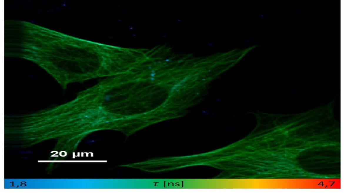
The fluorescence lifetime describes the average time of a fluorophore in its electronically excited state before it decays to the ground state. By coloring the image based on the statistical analysis of the extracted lifetimes, new information content can be revealed. The additional information can be used in many ways and is directly related to the sample (Oxygen amount, conformational change of enzymes) and its environment (pH-Value, temperature).
To overcome the difficulties in clinical applications that accompany a high priced module, the team of Thomas Hellerer will develop a cost-saving device for fluorescent lifetime imaging microscopy taking advantage of low-budget components, a clever setup and certain physical processes.

Running duration:
01.10.2019 - 30.09.2022
Funded by:
Österreichische Forschungsförderungsgesellschaft“ (FFG)
Project Partners:
Fachhochschule Vorarlberg GmbH
Prospective Instruments LK OG